Wound Healing Center


Orthopedic Services


Request Appointment
WOUND HEALING SPECIALISTS
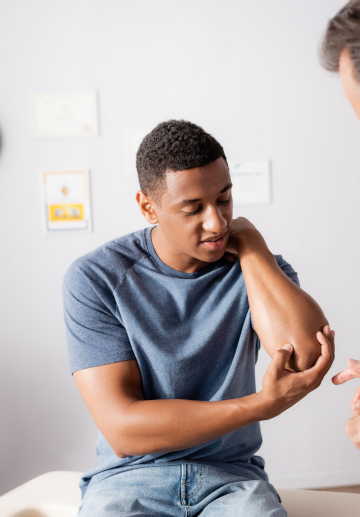
Our wound management starts with a thorough assessment of the wound and peri-wound skin.
Dr. Singh
One of our wound care treatment specialists will be the caregiver to assess changes in a patient’s skin and initiate wound care. Managing this can be challenging as patients present with diverse disorders and tissue damage can range from superficial to deep. But by applying a few basic principles starting with a skin and wound assessment our doctor can simplify the process and determine an appropriate treatment plan. The assessment should include the following components:
Wounds can be present over different anatomical parts of the body but the basic principles of choosing wound care bandages and dressings remain the same. Choosing the correct wound dressing treatment solutions will lessen wound healing time, provide cost-effective care and improve the patient’s quality of life.

VanTreia Gross, PA-C
The goal is to help the wound heal as soon as possible by using an appropriate dressing material to maintain the right amount of moisture. When the wound bed is dry use a dressing to increase moisture and if too wet and the surrounding skin is macerated use material that will absorb excess fluid and protect the surrounding healthy skin.
Critical criteria to consider before choosing a specific wound dressing are cleaning, absorbing and regulating. Choosing a dressing guided by cost, ease of application and clinician’s preference is essential. No need to wait for care for your wound in the hospital emergency room or clinic – reach out today for an appointment. Our wound experts can help you manage your wounds on an on-going basis to ensure proper healing happens.
ASSESSMENT BASICS
ANATOMIC LOCATION

DEGREE OF TISSUE DAMAGE
The degree of tissue damage in a wound can help guide its care plan and provide information about its healing trajectory. Wounds can be partial thickness (limited to the epidermal and/or dermal layers) or full-thickness (with damage in the subcutaneous layers and below). For pressure injuries, the National Pressure Ulcer Advisory Panel (NPUAP) staging classification is used to describe the appearance of the wound and extent of tissue damage..
TYPE OF TISSUE IN WOUND
The tissue in a wound bed can be viable or nonviable. Viable tissue may appear beefy red (granulation tissue) or light pink (new epithelial tissue). Nonviable or necrotic tissue varies in appearance: Eschar may be black, brown or tan; fibrin slough is stringy or adherent and yellow.
WOUND SIZE
Wound size can be described using linear dimensions (length times width). Measure the length along the head-toe axis and the width from side to side. If the wound has depth, measure from its deepest point to the surface using a sterile cotton-tip applicator.
WOUND EDGES AND PERI-WOUND SKIN
The outer edge of a wound can provide information about its duration and even its cause. Wounds over bony prominences with defined edges may be related to pressure. Venous wounds on the leg often have an irregular shape and undefined edges. Periwound skin can provide information about other factors that contribute to wound development or nonhealing. For example, weeping or excess drainage around a venous ulcer can macerate peri-wound skin, giving it a wet appearance that may be soft and gray-white.
INFECTION
During wound care assessment, note any signs or symptoms of local infection (erythema, induration, pain, edema, purulent exudate or odor). Keep in mind that patients with chronic wounds may not exhibit these classic signs due to biofilm presence. This extracellular polysaccharide matrix embeds microorganisms and delays healing.
PAIN
The presence and intensity of pain associated with a wound can provide important information about its cause and duration. However, pain level may not correlate with injury extent. Skin tears can be very painful because superficial skin damage exposes nerve endings in the dermal layer.
IDENTIFYING WOUND ETIOLOGY
PRESSURE INJURIES
Formerly known as pressure ulcers, these are defined by the NPUAP as localized damage to the skin and/or underlying soft tissue. They usually occur over a bony prominence or are related to medical or other devices. The injury can present as intact skin or an open ulcer and may be painful. It results from intense and/or prolonged pressure or pressure combined with shear. Soft tissue tolerance for pressure and shear may also be affected by microclimate, nutrition, perfusion, comorbidities and soft tissue condition. Pressure injuries are described using the NPUAP staging system based on clinically observed damage.
VENOUS ULCERS
These are related to valve incompetence in the lower extremities that allows blood to reflux into the superficial venous system causing edema. Incomplete emptying of deep veins can result in higher-than-normal peripheral venous system pressure in lower extremities which can eventually lead to ulcerations.
ARTERIAL WOUNDS
These result from severe tissue ischemia. One common cause of lower extremity arterial disease and ulceration is atherosclerosis of peripheral arterial vessels.
SKIN TEAR
A skin tear is defined by the International Skin Tear Advisory Panel (ISTAP) as a traumatic wound caused by mechanical forces (shear, friction or blunt force), such as removing adhesives. Severity may vary by depth but does not extend through the subcutaneous layer. The doctor should use ISTAP’s classification system to describe skin damage:
Type 1: no skin loss; a skin flap can be positioned to cover the exposed wound base.
Type 2: partial loss of the skin flap.
Type 3: total loss of the skin flap; the entire wound bed is exposed.
MASD
Moisture-Associated Skin Damage (MASD) is inflammation and erosion of skin due to exposure to various types of moisture such as urine, perspiration or wound drainage. Chronic exposure to moisture macerates skin, impairing its protective mechanisms and disrupting normal flora which can predispose patients to cutaneous infections like candidiasis. Incontinence-associated dermatitis (IAD), a subtype of MASD, is caused by chronic exposure to urine and/or liquid stool.
TOPICAL THERAPY
After assessing the wound and determining its cause, the doctor can initiate topical therapy. Topical dressings create an environment that fosters normal healing. Bryant and Nix outline eight objectives for caregivers to consider when selecting appropriate interventions.
PREVENTION & MANAGEMENT
A primary goal of topical wound care is to protect the wound base from outside contaminants like bacteria. If infection is evident in the wound, wound cultures should be considered and the need for topical antimicrobial/antiseptic products discussed with the primary provider.
Topical antibiotics destroy microorganisms while topical antiseptics inhibit microbial growth. Examples include cadexomer iodine, honey and silver sulfadiazine. These products are covered with a secondary dressing and can be used in partial-thickness and full-thickness wounds that are infected or at high risk for infection. They should not be used long-term.

CLEANSE THE WOUND
Routine cleaning should be performed at each dressing change using products physiologically compatible with wound tissue. Normal saline is least cytotoxic; when delivered at 4 to 15 PSI pressure it can remove wound debris. Commercially available wound cleansers may also be used but avoid hypochlorite solutions, betadine, hydrogen peroxide and acetic acid in routine cleansing as these agents can be cytotoxic to fibroblasts.
PRESSURE INJURIES
If necrotic tissue is visible in the wound bed, removal of this devitalized tissue is usually indicated. One exception is stable, dry eschar on the heel; in this case leaving the eschar in place until the patient’s vascular status can be determined is recommended.
Wound debridement can be accomplished using several methods. Autolytic debridement, the slowest form of debridement, uses moist topical dressings to foster autolysis of necrotic tissue. Enzymatic debridement involves applying a prescribed topical agent directly to the wound bed. It’s usually applied daily and covered with a dressing such as gauze or foam. Sharp wound debridement may be performed at bedside (conservative) or in an OR (surgical) by a qualified healthcare provider. Necrotic wounds showing signs of infection should be treated with sharp/surgical debridement as soon as feasible.
MAINTAIN APPROPRIATE MOISTURE
A moist wound environment facilitates healing, reduces pain and decreases infection risk. In heavily draining wounds, dressings that absorb excess drainage should be used to maintain appropriate moisture levels in the wound bed.
ELIMINATED DEAD SPACE
Deep wounds need to be packed with agents like normal saline or hydrogel-impregnated dressings to keep the wound bed moist. In excessively moist wounds alginate or Hydrofiber dressings can help control drainage. Packing material should be easy to remove during dressing changes to avoid injuring healing tissue.
CONTROL ODOR
To manage odor, if present, the nurse should consult with the provider about dressing change frequency, wound cleansing protocol and possible need for debridement or topical antimicrobials. The primary healthcare provider or wound care specialist should be consulted regarding treatment options to control wound odor.
PRESSURE INJURIES
Painful wounds should be thoroughly assessed for infection or other causes (such as an associated fracture or foreign object in the wound) and treated accordingly. Moisture-retentive dressings can decrease pain associated with dressing removal and reduce the need for frequent changes in painful wounds.
PROTECT PERI-WOUND SKIN
Heavily draining wounds or improper use of moist dressings can lead to peri-wound skin maceration which alters tissue tolerance and damages wound edges. Skin barrier creams/ointments, protective wipes or barrier wafers can protect peri-wound skin.
Selection of an appropriate topical dressing should be guided by these objectives. Always follow manufacturer guidelines as well as facility policies and procedures for specific product use.
WHOLE PICTURE
Since wounds do not occur in isolation, wound management involves not only topical care but also attention to other systemic or local factors that can contribute to wound development or impaired healing. Consider the following:
- In the case of pressure injuries, minimizing pressure and shear must be part of the treatment plan.
- For neuropathic wounds, decreasing further injury risk (especially in patients with diminished sensation) is an important educational intervention.
- Controlling lower extremity edema in patients with venous ulcers through compression is a fundamental component of successful treatment. Compression dressings and multilayer bandaging systems are gold standards for venous ulcers.
- Establishing adequate perfusion to extremities in patients with arterial ulcers is essential for wound healing. Some patients may need revascularization to support healing.
- In patients with skin tears, protecting skin is a priority (especially in those with fragile skin or other risk factors).
Nutrition is another component of an overall wound treatment plan. Many hospitalized patients may be malnourished. Proper nutrition and hydration are essential for normal cellular function and successful wound healing. It’s important to assess the patient’s nutritional status with the help of a registered dietitian to determine their potential to heal. A nutritional plan should consider the patient’s overall health and nutritional status and include appropriate supplementation.
Detailed documentation of ongoing assessment findings and interventions is an important communication tool for all caregivers, including the wound care professional who will clarify the plan of care.
Request an Appointment Online
Carolina Regional Orthopaedics, PA allows you to easily schedule an appointment online 24 hours a day, so you can continue your day with little disruption.
Our Providers
At Carolina Regional Orthopaedics, PA, all of our providers are Board Certified Orthopaedic Surgeons or

Bernard P. Kemker, MD

Glenn E. MacNichol, MD

Robert C. Martin, DO

Mark N. Perlmutter, MS, MD
Hardayal Singh, MD

Morgan Greene, PA-C
VanTreia M. Gross, PA-C
Paul Kelley, PA-C
William J. Mayo, PA-C
Vincent P. Keeney, PA-C
Joseph W. Williams, PA-C
Our Locations

Rocky Mount
Carolina Regional Orthopaedics, PA
Mon-Thurs
110 Patrick Ct, Rocky Mount, NC 27804
Hours:
Monday- Thursday: 8 AM - 6 PM
Closed
Friday - Sunday
Phone: 252-443-0400
Fax: 252-443-0572
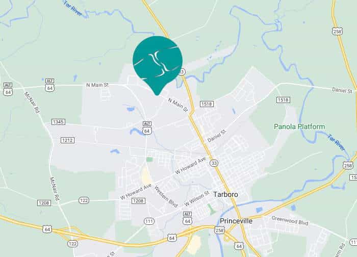
Tarboro
Carolina Regional Orthopaedics, PA
Tuesdays Only
Address:
101 Clinic Dr. Ste. 7A, Tarboro, NC 27886
Hours:
Tuesday: 8 AM - 5 PM
Closed:
Sunday - Monday, Wednesday - Saturday
Phone: 252-443-0400
Fax: 252-443-0572